Dr Mandeep Kaur
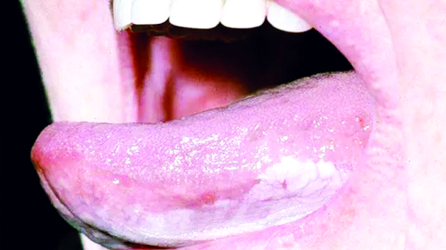
White patches or spots of the mouth are common and may be benign, potentially malignant or malignancy. Given the high-risk nature of some white patches, it is important to perform a thorough history and examination. For those routinely examining the oral mucosa, such as dentists, referrals should be in accordance with local guidance, others should refer to specialist centres.
Clinical examination
The site, size and distribution of the white patches.
Do the white patches wipe away?
Is there any associated ulceration or mucosal redness?
Is the overall appearance homogeneous or non-homogenous?
Local
Debris, burns (from heat, radiation, chemicals such as mouthwashes), grafts and scars may appear pale or white. Materia alba can usually easily be wiped off with a gauze.
Furred tongue: Tongue coating is common, particularly in edentulous adults on a soft, non-abrasive diet, poor oral hygiene, dehydration and those who have febrile diseases.
Traumatic
Frictional keratosis: Chronic abrasion of the mucosa by dentures, or rough edges of teeth or restorations.
Cheek/lip biting: Habitual biting of tongue or cheek triggering reactive areas of keratosis.
Stomatitis Nicotina : Thermal trauma from heavy smoking/pipe use triggers inflammation of minor salivary glands with hyperkeratosis of the surrounding mucosa especially hard palate.
Canker Sores: Single, large white patch in the mouth. It can look red around the white patch due to irritation from food or drink, tobacco use, injuries from accidentally biting the cheek or lip.
Infective
Fungal infections known as Candidiasis (thrush) is seen as soft raised white plaques which can be easily wiped off to reveal red underlying mucosa and is due to immunosuppressant drug use, following antibiotic therapy, or as a result of poor denture hygiene.
Hairy leukoplakia appears as painless, adherent, soft, white raised lesion with vertical corrugations caused by Epstein-Barr virus in those on immunosuppressant medications and late stage HIV infections.
Warts and papillomas (caused by human papillomaviruses)
Mucous patches and leukoplakia of syphilis. Specialist care is usually indicated.
Potentially malignant disorders
Leukoplakia: Well-defined, tough, adherent white plaques usually with a wrinkled, uneven surface. Common causes are tobacco chewing, cigarettes, pipe, cigar, snuff and reverse smoking (bidi). High risk sites are lateral border of tongue, floor of mouth and soft palate complex.
Oral submucous fibrosis: Pale, hard mucosa with palpable fibrous bands( fibrosis) most commonly involving cheek mucosa accompanied by limited mouth opening and dark brown stained teeth. Habitual chewing of paan, gutka, betel nut, slaked lime, nutritional deficiency are the common causes.
Oral lichen planus: White patch which may be linear, reticular, atrophic, erosive and occurs bilaterally on the buccal mucosa, related to diabetes, certain drugs, anxiety, stress, reactive dental fillings, hepatitis C infection, immunologically mediated, malaria.
Lupus erythematosus: Auto-immune connective tissue disease where the body produces autoantibodies against DNA, triggering inflammation. Discrete red and white areas sometimes with associated ulcers are seen.
Malignancy
Oral cancer: Known as squamous cell carcinoma is seen as exophytic whitish or ulcerative firm areas with rolled borders.
Underlying Health Conditions
Some health conditions that don’t have to do with your mouth can actually have white spots or sores in your mouth as a symptom.
Behcet’s syndrome
Celiac & Crohn’s disease
Ulcerative colitis
Normal variantss
Fordyce spots: Whitish or yellowish white spots on the buccal mucosa which represents ectopic sebaceous glands.
Leucoedema: Congenital whitish-grey filmy appearance especially of buccal mucosa, bilaterally which disappears if the mucosa is stretched.
Inherited dyskeratosis: White sponge nevus is seen as thickened, folded, soft white patches which fade into normal mucosa due to mutation in gene encoding keratin.
Management
For all new white patches that are not easily explained by trauma or normal anatomical variation, management should begin with a prompt referral for a specialist opinion. Biopsy is recommended in some cases to assist in attaining a definitive diagnosis.
Habit cessation and patient education
Replacement of fillings/denture if required
Treatment of acute infections
Appropriate referral
Topical corticosteroids
Immunomodulating drugs
Topical or systemic antifungals
Surgical Removal of the lesion
Regular monitoring of the oral mucosa
(The author is Assistant Professor Dept of Oral Pathology & Oral Microbiology Indira Gandhi Govt Dental College Jammu)


