Dr Arvind Kohli
Thoracic surgeons in current practice perform a variety of minimally invasive procedures for conditions of the lung and chest wall. Compared to surgery performed through a long, open-chest incision, minimally invasive lung surgery provides several important benefits for patients, which include:
Faster recovery and return to normal activities, shorter hospital stay, Less Pain Little scarring, Minimal bloods loss, No cutting of the ribs or breastbone (sternum) and Possible improves cure rates for cancer
At the Forefront of Minimally Invasive Thoracic Surgery
Following are the various techniques being practiced these days
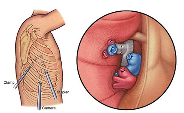
* Video-assisted thoracic surgery (VATS) involves insertion of a long, thin tube (called a thoracoscope) through a small incision, or port. The thoracoscope’s miniature camera allows the surgeon to view and examine the chest cavity. Additional specially designed instruments inserted through one or two more ports enable the surgeon to remove tissue. For more extensive operations, such as lung resection for cancer, an extra incision measuring about 5 centimeters is made for the removal of the lung tissue.
* Robotic thoracic surgery is performed using the da Vinci Surgical System. This sophisticated robotic approach, like VATS, gives the surgeon access inside the chest cavity through tiny incisions. The surgeon controls the robot’s movements from a nearby console in the operating room. The robotic system provides improved visualization (using three-dimensional technology), better access to mediastinal tissues, and improved ability to remove lymph nodes as part of a cancer operation.
The use of these techniques is evaluated on a case-by-case basis. Our surgeons choose the best approach for each patient, taking into account many factors, such as the patient’s particular condition, medical history and anatomy.
Whenever possible, thoracic surgeons regularly use minimally invasive techniques to remove nodules and tumors for an accurate and swift diagnosis. Lymph nodes in the chest are also biopsied to determine the stage of the cancer. If cancer is confirmed, the surgeon may perform one of the following procedures using VATS or robot-assisted techniques:
* Wedge resection: Removal of the tumor and tissue surrounding the cancer.
* Segmental resection: Removal of the tumor and surrounding anatomic structures consistent with a more complete cancer operation. This may be appropriate for very small, peripheral tumors.
* Lobectomy: Removal of the entire lobe of the lung that contains the cancerous tissue. This is the standard operation for most lung cancers. More than half of our lung resection operations are performed using minimally invasive techniques
Pleural Effusion
Pleural effusion is the presence of excess fluid in the pleural space, the area between the lungs and the chest wall. VATS surgery may be performed to remedy this problem by: removing restricting tissue around the lung; applying medicine to reduce fluid accumulation; or inserting a temporary drainage tube, which substantially reduces symptoms associated with the fluid collection.
Pneumothorax
Pneumothorax occurs when air leaks into the space between the lungs and chest wall, causing part or all of a lung to collapse. VATS surgery is performed to prevent recurrence of the problem, or when prior treatment with a chest tube is unsuccessful. Any abnormal blisters on the surface of the lung that contribute to air leakage are removed during the procedure. In addition, medicine is applied to the pleural surfaces to reduce the risk of future lung collapse.
Esophageal Cancer
Patients who have esophageal cancer and cancer of the junction between the esophagus and stomach are often candidates for resection of the esophagus. Surgeons performs resections in most patients using a minimally invasive approach for both the abdominal and the thoracic portions of the operation. (In most cases, patients who have these procedures have undergone preoperative chemotherapy or chemotherapy combined with radiation therapy.) This technique may reduce the risk of complications after surgery, shorten hospital stay, and provide equivalent long-term benefits for cancer survival.
Mediastinal Tumors
Mediastinal tumors are tumors that develop in the mediastinum — the area of the chest that separates the lungs and contains the heart, aorta, esophagus and trachea. These tumors can be seen in the front (anterior), middle, or back (posterior) of the mediastinum. They form and grow in the thymic (thymoma), nerve (neural), lymphatic (lymphoma) or soft tissue. Although open surgery is sometimes necessary, our thoracic surgeons use VATS or robot-assisted techniques to remove mediastinal tumors whenever possible.
Thymectomy for myasthenia gravis: Thymomas (mediastinal tumors seen in the thymus) are sometimes found in people diagnosed with myasthenia gravis. Thymectomy, or surgical removal of the thymus, is sometimes appropriate for patients with myasthenia gravis – even when no tumor is present – as a means of improving their neurologic symptoms.
Other Conditions
In addition to the conditions listed above, our thoracic surgeons perform minimally invasive surgery for the following conditions:
* Achalasia, a disorder of the esophagus that makes it difficult to swallow food.
* Barrett’s esophagus, a precancerous condition that develops as a result of gastroesphageal reflux disease.
* Gastroesophageal reflux disease (GERD), a condition when a higher-than-normal amount of gastric juice refluxes from the stomach back into the esophagus.
* Hyperhidrosis, or excessive sweating. Using endoscopic techniques, surgeons can destroy or remove the sympathetic nerve, which greatly reduces symptoms.
(The author is a Heart Surgeon at GMC Jammu)
Trending Now
E-Paper


