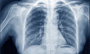
NEW YORK: In a first, researchers have described the chest X-ray scan features that may aid in the early detection and diagnosis of the novel Chinese coronavirus which has so far killed nearly 500 people in China.
According to the World Health Organization (WHO), the virus that has been temporarily named, the “novel coronavirus” (2019-nCoV) causes respiratory illness resembling viral pneumonia, resulting in fever, cough, and shortness of breath.
The current study, published in the journal Radiology, characterised the key X-ray imaging findings in chest CT scans in a group of 21 patients infected with 2019-nCoV in China.
The patients consisted of 13 men and 8 women ranging in age from 29 to 77 years old, with a mean age of 51.2 years.
According to the researchers, including those from the Mount Sinai Health System in the US, the initial CT scans of the patients were evaluated for several chest features.
These included the presence of partial filling of air spaces in the lungs, liquid deposition in air sacks, and the number of lung lobes affected by these discrepancies.
The researchers also assessed the chest scan images for the presence of nodules, liquid discharge, abnormal lymph node size, and underlying diseases like fibrosis.
According to the study, the 2019-nCoV typically manifests on CT scans which show air sack liquid deposition, and filling up of the air spaces. (AGENCIES)


