Dr Arvind Kohli
Heart is one of the most important organs of our cardiovascular and circulatory system and if there is even a minute problem in its structure of functioning it indeed is a grave thing. Heart attacks, blockages are a common thing. But a heart murmur of having a hole in the heart is certainly very serious and which cannot be prevented as it is a natural defect in almost all the cases. Many young infants are born with such type of problems. But due to clinical advancements, now such problems can be fixed and new lease of life is given to the patient.
What is a hole in heart?
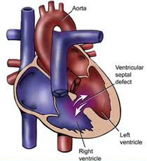
It is a very simple type of congenital heart defect. Hole in the heart is a problem related to the structure of the heart and is present right from the birth. Due to this defect in the heart, the normal blood flow is changed. The heart cornprises of two sides which are separated by septum-an inner wall. With every beat of the heart, its right side gets oxygen deficient blood from the body and directs it through pumping towards the lungs. Similarly, the heart’s left side gets oxygen rich blood via lungs and then pumps into the body from where it is reached to various cells units of different organs. The key role here is of the septum which divides the two sides and stops the blood from two sides mixing with each other.But sometimes babies are born with a small hole in the lower or upper part of the septum. When the hole is present in the upper two chambers (atria) of the septum it is known as atrial septal defect or ASD. When the hole is in the septum in between the lower two chambers of the heart (ventricles), the defect is termed as ventricular septal defect (VSD).
What Causes Holes in the Heart?
Mothers of children who are born with atrial septal defects (ASDs), ventricular septal defects (VSDs), or other heart defects may think they did something wrong during their pregnancies. However, most of the time, doctors don’t know why congenital heart defects occur. Heredity may play a role in some heart defects. For example, a parent who has a congenital heart defect is slightly more likely than other people to have a child who has the problem. Very rarely, more than one child in a family is born with a heart defect.Children who have genetic disorders, such as Down syndrome, often have congenital heart defects. Half of all babies who have Down syndrome have congenital heart defects.Smoking during pregnancy also has been linked to several congenital heart defects, including septal defects.Scientists continue to search for the causes of congenital heart defects.
Signs and Symptoms
Atrial Septal Defect: Many babies who are born with atrial septal defects (ASDs) have no signs or symptoms. However, as they grow, these children may be small for their age. When signs and symptoms do occur, a heart murmur is the most common. Often,a heartmurmur is the only sign of an ASD..However a child presents with features of
Fatigue (tiredness)
Tiring easily during physical activity
Shortness of breath
Crowth stunting
Ventricular Septal Defect :
Babies born with ventricular septal defects (VSDs) usually have heart murmurs. Murmurs may be the first and only sign of a VSD. Heart murmurs often are present right after birth in many infants. However, the murmurs may not be heard until the babies are 6 to 8 weeks old.Most newborns who have small VSDs don’t have heart-related symptoms. However, babies who have medium or large VSDs can develop heart failure. Signs and symptoms of heart failure usually occur during the baby’s first 2 months of life.The signs and symptoms of heart failure due to VSD are similar to those listed above for ASD, but they occur in infancy.A major sign of heart failure in infancy is poor feeding and growth. VSD signs and symptoms are rare after infancy. This is because the defects either decrease, in size,close on their own or they’re repaired.
Diagnosing Hole in Heart If your child is discovered to have a heart murmur, in addition to doing a physical exam, the cardiologist take your child’s medical history. If a ASD. VSD is suspected, one or more of these tests:shall be dignostic
*Chest X-ray and electrocardiogram (EKG)’
*An echocardiogram (echo), which uses sound waves to produce a picture of the heart and to visualize blood flow through the heart chambers. This is often the primary tool used to diagnose a VSD.
A cardiac catheterization, which provides information about the heart structures as well as blood pressure and blood oxygen levels within the heart chambers. This test is usually performed for VSD only when additional information is needed that other tests can
What is the treatment for ASD and VSD?
Treating an Atrial Septal Defect
If a child has an atrial septal defect (ASD), routine checkups are done to see whether it closes on its own. About half of all ASDs close on their own over time, and about 20 percent close within the first year of life. lf an ASD requires treatment, catheter or surgical procedures are used to close the hole. Doctors often decide to close ASDs in children who still have medium-or large-sized holes by the time they’re 2 to 5 years old.
Surgery:
Open-heart surgery generally is done to repair secundum premium or sinus venosus ASDs. Then, the cardiac surgeon makes an incision (cut) in the chest to reach the ASD. Procedure involves repairing the defect with a special patch that covers the hole. A heart-lung bypass machine is used during the surgery so the surgeon can open the heart. The machine takes over the heart’s pumping action and moves blood away from the heart. The outlook for children who have ASD surgery is excellent. On average, children spend 3 to 4 days in the hospital before going home. Complications, such as bleeding and infection, are very rare.
Catheter Procedure a catheter (a thin, flexible tube) is inserted into a vein in the groin (upper thigh) and threaded the tube to the heart’s septum. A device made up of two small disks or an umbrella-like device is attached to the catheter.When the catheter reaches the septum, the device is pushed out of the catheter. The device is placed so that it plugs the hole between the atria. It’s secured in place and the catheter is withdrawn from the body.Within 6 months, normal tissue grows in and over the device.
The closure device does not need to be replaced as the child grows.
Treating a Ventricular Septal Defect
More than half of VSDs eventually close, usually by the time children are in preschool. Checkups may range from once a month to once every 1 or 2 years. If treatment for a VSD is required, options include extra nutrition and surgery to close the VSD. We can use catheter procedures to close some VSDs. We may use this approach if surgery isn’t possible. More research is needed to find out the risks and benefits of using catheter procedures to treat VSDs.
Surgery
Most doctors recommend surgery to close large VSDs that are causing symptoms, affecting the aortic valve, or haven’t closed by the time children are 1and a half year old. Surgery may be needed earlier if:
* A child doesn’t gain weight > Failure to thrive
Intractable congestive heart failure
Medicines are needed to control the symptoms of heart failure
Rarely, medium-sized VSDs that are causing enlarged heart chambers are treated with surgery after infancy. However, most VSDs that require surgery are repaired after first year of life. Doctors use open-heart surgery and dacron patches to close VSDs
Living With Holes in the Heart
The outlook for children who have atrial septal defects (ASDs) or ventricular septal defects (VSDs) is excellent. Advances in treatment allow most children who have these heart defects to live normal, active lives with no decrease in lifespan.
Many children who have these defects need no special care or only occasional checkups with a cardiologist (a heart specialist) as they go through life.
Trending Now
E-Paper


